The human skeletal system serves as the framework for the body, providing structure, support, and protection. It’s composed of 206 bones (in the adult human) that vary in size and shape, and are categorized into two main groups: the axial skeleton and the appendicular skeleton.
- Axial Skeleton: This includes the skull, vertebral column, and rib cage. It primarily provides protection for the body’s vital organs.
- Appendicular Skeleton: This includes the bones of the arms, legs, pelvis, and shoulder area. It facilitates movement and supports the axial skeleton.
Directions:
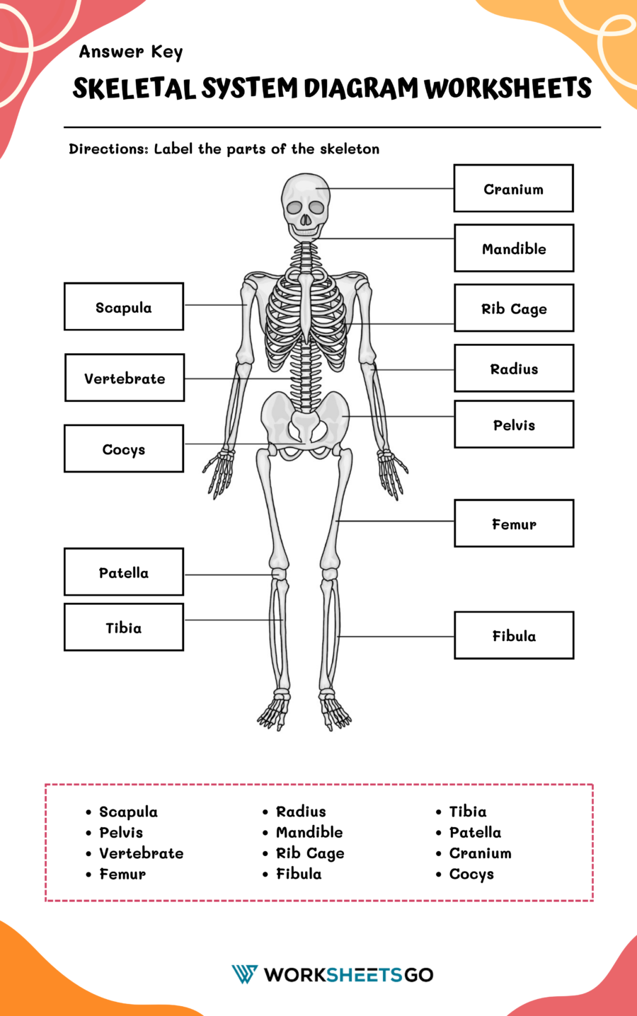
Label the following parts of the skeleton on your diagram worksheet. Use the provided list of bone names and locate each one on the diagram of the human skeleton.
List of Bones to Label:
- Skull:
- Cranium
- Mandible (Jawbone)
- Vertebral Column (Spine):
- Cervical Vertebrae
- Thoracic Vertebrae
- Lumbar Vertebrae
- Sacrum
- Coccyx (Tailbone)
- Rib Cage:
- Ribs
- Sternum
- Upper Limb:
- Humerus (Upper Arm)
- Radius and Ulna (Forearm)
- Carpals (Wrist)
- Metacarpals (Hand)
- Phalanges (Fingers)
- Pelvic Girdle:
- Ilium
- Ischium
- Pubis
- Lower Limb:
- Femur (Thigh)
- Tibia and Fibula (Lower Leg)
- Patella (Kneecap)
- Tarsals (Ankle)
- Metatarsals (Foot)
- Phalanges (Toes)
Worksheet Activity:
- Utilize a skeleton diagram worksheet that includes a clear, unlabeled illustration of the human skeleton.
- On your worksheet, carefully write the name of each bone (from the list above) beside the corresponding bone on the diagram. You might want to use arrows to indicate precisely which bone you’re labeling if the diagram is detailed or smaller in size.
- Ensure that the labels are clear and legible, making it easy to review and reference in the future.

Answer Key


